Prepare for tomorrow, today πŸ‚

Your last chance!

Register to our newsletter now and be informed when you should start to prepare for tomorrow!
This is it – if you don't gather your nuts of knowledge now, you will run into problems tomorrow! Your last opportunity to secure access to a stock of knowledge with -30% off plus Lifetime Access to all our courses ends on October 26th!

0+

Alumni

00

Days

00

Hours

00

Minutes

00

Seconds

Want to check whether you know the correct answer to our quiz?

Echo Icon

Gather your skills in echocardiography​!

Stockpile your knowledge 🌰 and thrive in your career with our echo courses.

Special Preparation Package with Guidelines in Focus

With our course Guidelines in Focus: Echo Essentials for Clinical Practice, be prepared for tomorrow. Secure your nuts of knowledge with this special fall deal!

Monthly subscription

89 €

instead of 99 €

Minimum of 3 Months Run-Time
10% Discount
Incl. live Q&As with Prof. Binder (next webinar on Oct. 30th!)
New content every week
CME credits
Incl. AI SonoAssistant access
Purchase
6 Months Access

349 €

instead of 499  €

6 Months Access
30% Discount
Incl. live Q&As with Prof. Binder (next webinar on Oct. 30th!)
New content every week
CME credits
Incl. AI SonoAssistant access
Purchase
12 Months Access

559 €

instead of 699  €

12 Months Access
20% Discount
Incl. live Q&As with Prof. Binder (next webinar on Oct. 30th!)
New content every week
CME credits
Incl. AI SonoAssistant access
Purchase

Build your expertise and benefit until October 19th from:

30% Discount 
​

Available on all our courses!

Lifetime Access
​

No expiration date on your course!

30 Day Money-Back Guarantee

No questions asked. Max. 30% course progress.

Echo Icon

Prepare now to build your foundation!

Are you nuts for knowledge? πŸ₯œ Our courses help gather your skills to survive the winter!

Echo Icon

Stock up on your MSK ultrasound knowledge!

We'll keep you out of the rabbit hole and show you what you really need to know πŸ’‘

Emergency Icon

Gather your skills in emergency and critical care before the cold hits!

Our courses give you the skills and expertise you need to thrive in your success – prepare now to succeed later.

30 Day Money-Back Guarante

We offer a 30 day money-back guarantee. 100% refund, no questions asked. Course progress must be under 30%.

Top rated by

Google

Trustpilot

Why our users love us

Find our ultrasound quiz questions, and answers here​!

Discoveries in the Ultrasound Jungle 🌴

Examine the following ultrasound images labeled A, B, and C. 

Based on your observations, how would you describe your findings and what would be the most probable diagnosis based on these images only? Make your diagnosis and then check for the answer!

And here is the correct answer!

What are the most probably diagnoses?

Click here for the answer!

Image A) This ultrasound image shows a diffusely heterogeneous thyroid gland. The parenchyma appears hypoechoic with coarse echotexture. What is the most likely diagnosis based on this ultrasound appearance?
πŸ‘‰ Hashimoto's Thyroiditis

Image B) This ultrasound image depicts an enlarged thyroid gland with multiple confluent nodules of varying size. They appear as heterogeneous masses within the thyroid tissue. The nodules also have cystic components. What condition is illustrated in this ultrasound image?
πŸ‘‰ (Multi)nodular Goiter

Image C) This ultrasound image shows a well-circumscribed, oval-shaped structure in the thyroid region. The structure has a hypoechoic cortex and a central echogenic hilum, typical of a lymph node. What is the structure shown in this ultrasound image?
πŸ‘‰ Lymph Node in the Thyroid Region

And if you want to learn to how to see the clues in the jungle of ultrasound, get our Thyroid Ultrasound MasterClass now with -30% plus Lifetime Access!

Want to test more skills or check the answer to previous cases? Check below πŸ‘‡

More Echo quizzes

OMG! What is this in the heart? 😱

A glimpse inside the heart. Something enters. Can you tell what it is? Do you need to worry?

  • What's inside the heart? *

And here is the correct answer!

What's inside the heart?

Click here for the answer!

The correct answer is B: Needle.

What you saw wasn’t a mystery – it was precision at work.

This is a transseptal puncture needle, captured during echocardiographic guidance of a TEER procedure.

Did you see it right away or was it hidden in the shadows of the ultrasound jungle? Let us guide you through with -30% off plus Lifetime Access to our courses!

Lost in the apex – what’s your next move?

You’re deep in the apical view, but the landscape looks unfamiliar. The apex doesn’t behave the way it should – compact, quiet, almost hiding something. In the jungle of ultrasound, some findings don’t roar – they whisper. 🀫

But what should catch your attention? Sometimes, key findings are hidden deep in the shadows. You’re the guide now: which imaging step will lead you out of the thicket and closer to the truth? πŸ‘€

  • What would you do? *

And here is the correct answer!

What would you do next?

Click here for the answer!

The correct answer is B: Contrast-enhanced echocardiography.

Apical HCM often evades detection on standard 2D imaging due to poor apical definition. A contrast study outlines the true cavity shape – the classic β€œspade-like” configuration that confirms the diagnosis. This subtle but crucial finding reminds us that even in familiar views, pathology can remain hidden. In the jungle of cardiac imaging, contrast is often the torch that reveals the path. Let us guide you through the jungle of ultrasound – with -30% off plus Lifetime Access to our courses!

Can you detect the abnormality in this "apparently normal" short axis view?

Can you spot the abnormal? Looks like a normal SAX view – or is it?

In the jungle, some things hide in plain sight.

Can you spot what’s camouflaged here? 🌴

  • Which structure is abnormal? *

And here is the correct answer!

Can you detect the abnormality in this "apparently normal" short axis view?

Click here for the answer!

Caught in the echo jungle: a rare 3rd papillary muscle!

 Even in the heart’s jungle, some things hide well.

This rare variant shows a third papillary muscle – a reminder that papillary anatomy can vary in size, length, location, and even number.

What you spotted is subtle – and rare.

This case shows a third papillary muscle in the left ventricle, an uncommon anatomical variant. 🧠 The papillary muscles can vary greatly in: Location, Size, Length; and, as seen here – even number. While often benign, such variations can sometimes be linked to mitral valve abnormalities or LVOT dynamics.

Always worth a closer look!

Did you answer this quiz correctly or do you need help spotting such hidden pathologies? Our expert adventurers can help you – our courses are now available with -30% plus Lifetime Access!
More ultrasound quizzes

A deadly embolism in a 20-year-old?! You won’t believe what we found…

A 20-year-old male from rural Romania presented with acute peripheral limb ischemia due to complete occlusion of the right femoropopliteal artery. His mental status was altered – he showed lethargy and very slow responsiveness.

A thrombectomy was performed. The extracted material did not resemble typical thromboembolic debris; instead, it was made up of amorphous, lamellar, shell-like structures. Was this really a classic thromboembolic event? These findings together with the young age and the absence of any risk factors prompted an echo exam – look at these incredible images... What differential diagnoses come to your mind?

  • What differential diagnoses come to mind? *

And here is the correct answer!

Which differential diagnoses come to your mind? πŸ‘‡

Click here for the answer!

The correct answer is B!

The patient had a primary cardiac manifestation of Echinococcus cysticus.

Treatment: Antihelminthic therapy with albendazole and praziquantel & surgical removal of the cardiac hydatide cyst + mitral valve replacement.

But unfortunately, the patient's condition further deteriorated... A follow up post is coming soon!

And if you want to learn to solve these cases in the jungle of ultrasound, get our courses now with -30% plus Lifetime Access!

Which pathology does the β€œsnake sign” describe? 🐍

Are you good with reptiles?

  • Which pathology does the snake sign describe? *

And here is the correct answer!

Which pathology does the β€œsnake sign” describe? 🐍

Click here for the answer!

The correct answer is C!

Thrombus in the right atrium originating from the IVC or femoral veins!


The snake sign or snake-like thrombus is clearly a frightening pathology. Its name however comes from its appearance, which resembles that of a snake. The thrombus forms in the veins of the lower extremity or IVC and therefore has the shape of a snake. If it dislodges it will cause pulmonary embolism (or even worse paradox embolism). Along its path it is often caught in the right heart where it can be detected with echo. The following image shows such a snake thrombus in a transthoracic subcostal view.

Could you see this hidden pathology or do you still need help? We teach you how to detect such pathologies – and our courses are now available with -30% plus Lifetime Access!
More MSK ultrasound quizzes

🌞 Running wild this summer? Don’t let this pathology slow you down!

πŸ” Check out this MSK ultrasound image highlighting a pathology often causing lateral knee pain in runners and cyclists. MSK ultrasound allows straight-forward diagnosis  to make your patients fit again for a sporty summer. 

πŸ“Œ Of course, proper MSK ultrasound requires scanning in two planes – but here we’ve only got the still image of a long-axis view of the structure in question. Based on the sonoanatomy and visible pathology, you should be able to make a well-informed guess... πŸ‘€

And here is the correct answer!

What's the most likely diagnosis? πŸ‘‡

Correct answer and explanation

The correct answer is B!

This long-axis MSK ultrasound image shows thickening and hypoechoic fluid collection in the area of the iliotibial band (ITB) as it passes over the lateral femoral condyle – a hallmark of ITB friction syndrome, commonly also called "runner’s knee."

πŸ”The ITB does not appear thickened but here is hypoechoic fluid/ edema deep to the band.

The location (lateral knee) and the sonopathologic caracteristics point to ITB pathology, not a medial meniscal tear (A) or a posterior popliteal cyst (C)!

And if you want to learn to solve these cases in the jungle of ultrasound, get our courses now with -30% plus Lifetime Access!
More Critical Care ultrasound quizzes

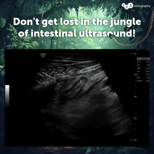
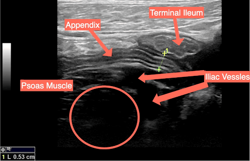
Don't get lost in the jungle of intestinal ultrasound!

A 36-year-old man came in with abdominal pain in the right lower quadrant, and he has no fever. Can you crack the case?

  • Which landmark do you use when scanning this area? *

And here is the correct answer!

Which landmark do you use when scanning this area?

Click here for the answer!

The correct answer is E) All of the above!

All of the above are correct! When scanning for the appendix, key landmarks like the iliac vessels, psoas muscle, terminal ileum, and ascending colon are crucial for orientation. However, finding a healthy appendix is tough, with only 30% being locatable due to its elusive nature in ultrasound scans.

Learn to solve such emergency cases in an instant with our Prehospital Point-of-Care Ultrasound course! Now available with -30% plus Lifetime Access!

00

Days

00

Hours

00

Minutes

00

Seconds

Are you busy gathering your skills?

We make it easy for you to build a strong foundation to survive the winter!
πŸ‘‡ All courses available!

30% Discount 
​

Available on all our courses!

Lifetime Access
​

No expiration date on your course!

30 Day Money-Back Guarantee

No questions asked. Max. 30% course progress.

This offer ends for good on October 26th, 2025.