Hatch your echocardiography skills!
See what's inside the heart with echocardiography and don't chicken out of learning!
Echo MasterClass
43.5 CME credits (more coming!)
Advanced echocardiography
instead of 1,470 €
only 1,029 €
TEE MasterClass
11 CME credits
Transesophageal echocardiography
instead of 880 €
only 616 €

Echo BachelorClass
12.75 CME credits
Basic echocardiography
instead of 830 €
only 581 €
Speckle Tracking MasterClass
12.25 CME credits
Deep knowledge of strain rate imaging
instead of 940 €
only 658 €

Cardiac Filling MasterClass
6 CME credits
Advanced echocardiography
instead of 830 €
only 581 €
Right Heart MasterClass
7.25 CME credits
Right heart clinical course
instead of 770 €
only 539 €
ACHD BachelorClass
16.5 CME credits
Basics of common and (more or less) simple adult congenital heart defects
instead of 940 €
only 658 €
HCQ MasterClass
4.75 CME credits
Guidelines of the quantification of heart chambers and walls
instead of 590 €
only 413 €
Carotid MasterClass
10.5 CME credits
Advanced carotid and vertebral artery ultrasound
instead of 930 €
only 651 €
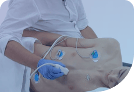
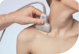
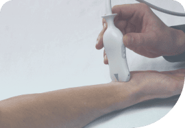
See what's inside with ultrasound!
We help you see what's inside with ultrasound! Hatch your skills now – swipe for even more courses.
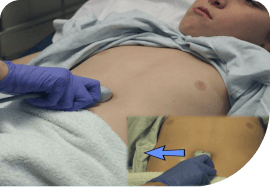
Abdominal US BachelorClass
10 CME credits
Detect abdominal pathologies with ultrasound
instead of 770 €
only 539 €
Thyroid US MasterClass
10.5 CME credits
Basics and advanced techniques of thyroid ultrasound
instead of 830 €
only 581 €
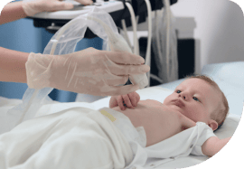
Neonatal & Pediatric US BachelorClass
4.75 CME credits
Ultrasound knowledge and skills specific to neonates and infants
instead of 830 €
only 581 €

Lung Ultrasound MasterClass
6.5 CME credits
Basic and advanced lung ultrasound
instead of 750 €
only 525 €
Vascular Lower Extremity BachelorClass
9.5 CME credits
Non-invasive vascular ultrasound
instead of 770 €
only 539 €
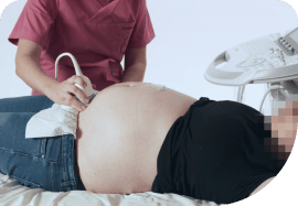
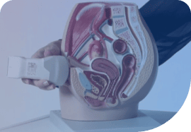
OB/GYN Ultrasound BachelorClass
7 CME credits
Basics of gynecology & obstetrics
instead of 840 €
only 588 €
Ultrasound Guided Regional Anaesthesia
Expertise in regional anaesthesia techniques
instead of 700 €
only 490 €
Pediatric Ultrasound BachelorClass
4.75 CME credits
Diagnose pediatric patients through ultrasound
instead of 770 €
only 539 €
OB/GYN POCUS FocusClass
2 CME credits
Knowledge and skills on how to do an ultrasound examination in OB/GYN
instead of 420 €
only 294 €
We help you see what's inside with MSK ultrasound!
Hatch your skills in musculoskeletal ultrasound so you can see what's inside! Swipe for more courses!
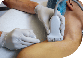
Musculoskeletal Ultrasound BachelorClass
19.75 CME credits
Foundations of MSK ultrasound
instead of 990 €
only 693 €

MSK Guided Injections US MasterClass
4.75 CME credits
Advanced MSK ultrasound
instead of 770 €
only 539 €
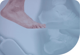
MSK Foot & Ankle Ultrasound BachelorClass
11.5 CME credits
Basics of MSK US of the foot and ankle joint
instead of 700 €
only 490 €
Bone Fracture FocusClass
2.5 CME credits
Ultrasound knowledge and skills specific to fracture sonography
instead of 300 €
only 210 €
Neuromuscular US Essentials
8.5 CME credits
Diagnose conditions and injuries not visible through other imaging methods
instead of 630 €
only 441 €
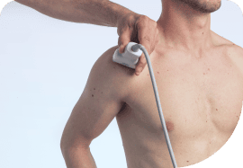
MSK Ultrasound of the Shoulder Joint
20 European CME credits
Ultrasound knowledge and skills specific to shoulder joint sonography
instead of 820 €
only 574 €

Hatch your ultrasound skills for emergencies and point-of-care ultrasound!
Be prepared for emergencies at all times – we help you see what's inside with ultrasound! Don't chicken out of learning.
Point-of-Care US FocusClass
6 CME credits
Primary care ultrasound
instead of 830 €
only 581 €
Emergency & Critical Care US Essentials
Essentials of emergency ultrasound
instead of 790 €
only 553 €
Prehospital Point-of-Care Ultrasound
8 CME credits (more to come!)
POCUS in prehospital scenarios (2 chapters to be published)
instead of 1,030 €
only 721 €
Emergency US MasterClass
5.25 CME credits
Advanced emergency ultrasound
instead of 770 €
only 539 €

Emergency US BachelorClass
Basic emergency ultrasound
instead of 700 €
only 490 €
Why our users love us

Ahmed Benzoghli

Wasseem Rock

Kati Tonninger, Austria
"I was recommended 123sonography through a friend and became a fan. The quality of the images and films are amazing."

Steven Feldstein, USA
"The e-learning platform is quite simple to navigate through, and the quality of the videos and images is quite good."

Satish Govind, India
"The communication style is simple, uncomplicated, but extremely effective in the way various topics are covered and discussed."
Find our quiz questions and answers here!
Clinical Reasoning in Action 🔍
Which of the following statements about pulmonary embolism is true?
A) The most specific ECG finding is ST-elevation in V2–V4
B) Tachycardia is the most common ECG abnormality in PE
C) Pulmonary embolism usually presents with bradycardia and normal oxygen saturatio
D) Prior asthma or COPD is the strongest independent risk factor for P
E) S1Q3T3 on ECG is both highly sensitive and specific for diagnosing PE
And here is the correct answer!
Which of the following statements about pulmonary embolism is true?
Fruits and heart disease – what’s the connection? 🍎💓
Which fruit do you associate with HCMP?
And here is the correct answer!
Which fruit do you associate with HCMP?
Are you an echo Guru? 👀
Which patient has an anterior WMA?
And here is the correct answer!
Which patient has an anterior WMA?
Not all bicuspid valves are equal 💡
Which of these bicuspid valves is most common?
And here is the correct answer!
Which of these bicuspid valves is most common?
Brain Ultrasound Case 🔍
Which of the following statements about choroid plexus cysts on ultrasound and the use of color Doppler is TRUE?
A. B-Mode and color Doppler are not sufficient - In any case CT is required to confirm the diagnosis
B. Typical choroid plexus cysts are anechoic, round or oval‑shaped structures with no internal blood flow on color Doppler
C. Cysts with septations or echogenic components may suggest other etiologies—such as prior hemorrhage or vascular malformations—and warrant further evaluation
D. It is most important to identify calcifications within the cyst(s)
E. Color Doppler is used to detect active hemorrhage into the cyst(s)
And here is the correct answer!
Which of the following statements about choroid plexus cysts on ultrasound and the use of color Doppler is TRUE?
🥚 What Do These Strange Eggs Have to Do with the Heart?
Which image do you associate with dilated CMP?
And here is the correct answer!
Which image do you associate with dilated CMP?
When evaluating for a partial-thickness tear of the Achilles tendon on ultrasound, which of the following findings are suggestive of this diagnosis?
A. The paratenon tissue is taking up the space of the tendonB. Hypoechoic defect within otherwise continuous tendon fibersC. Focal compressibility of a well-defined hypoechoic gapD. Absence of fibrillar pattern and continuity
And here is the correct answer!
When evaluating for a partial-thickness tear of the Achilles tendon on ultrasound, which of the following findings are suggestive of this diagnosis?
Can you "eye ball" dyssynchrony?
Which patient(s) have an LBBB?
And here is the correct answer!
Which patient(s) have an LBBB?
Can you spot the clot? 🩺🌷
And here is the correct answer!
Can you spot the clot? 🩺🌷
Which of the following do you associate with a specific pattern in cardiology?
A) Tail of a cat 🐈⬛
B) Chicken legs 🐓C) Rabbit ears 🐰D) Parrot head 🦜
And here is the correct answer!
Which of the following do you associate with a specific pattern in cardiology?
🌸 Spring, Blooms & Bleeding – Can You Spot the Blood?
And here is the correct answer!
Which of these structures is most likely to contain blood?
Can you solve this quiz about hydronephrosis?
Which of the following ultrasound findings are used to grade the severity of hydronephrosis?
A) Degree of dilation of the renal pelvis and calycesB) Presence of perinephric fat strandingC) Thinning of the renal cortexD) Regularity /Irregularity of the kidney tissue surfaceE) Visibility and separation of calyceal structures
And here is the correct answer!
Which of the following ultrasound findings are used to grade the severity of hydronephrosis?
🌸 Which Spring Symbol Fits This Condition?
Which image do you associate with Libman-Sacks endocarditis?
And here is the correct answer!
Which image do you associate with Libman-Sacks endocarditis?
Learn MSK Ultrasound and Injection Techniques – Essential Skills for Your Practice!
In which situation does a corticosteroid injection make the most sense for treating tennis elbow?
And here is the correct answer!
In which situation does a corticosteroid injection make the most sense for treating tennis elbow?
Spring, Eggs &… Calcifications? 👀
Which of these pathologies is termed egg shell calcifications?
And here is the correct answer!
Which of these pathologies is termed egg shell calcifications?
Do you know what's so different about performing and interpreting ultrasound exams on our youngest patients?
Which of the following statements about the difference between neonatal and adult lung ultrasound diagnostics is WRONG?
And here is the correct answer!
Which of the following statements about the difference between neonatal and adult lung ultrasound diagnostics is WRONG?
🥚 Spot the Rotten Egg – Which One Is Malignant?
And here is the correct answer!
🥚 Spot the Rotten Egg – Which One Is Malignant?
Are eggs healthy or not? 🥚
Which condition is less likely in individuals with moderate egg consumption?
And here is the correct answer!
Which condition is less likely in individuals with moderate egg consumption?
Which animal’s heart has four chambers?
Which animal's heart has four chambers?
A) Chicken
B) Fish
C) Frog
D) Lizard
And here is the correct answer!
Which animal's heart has four chambers?
Can you spot twins on an early pregnancy scan?
What do the Lamda- and T-sign refer to in OB/GYN ultrasound?
Made your guess? Then check if you are right here 👇
Hearts and… cacti? 🌵 Chickens? 🐔 Cauliflower? 🥦 What’s going on here?
Which morphology of the left atrial appendage has the highest thromboembolic risk?
A) Windsock
B) Chicken wing
C) Cauliflower
D) Cactus
And here is the correct answer!
Which morphology of the left atrial appendage has the highest thromboembolic risk?
00
Days
00
Hours
00
Minutes
00
Seconds
Don't chicken out of learning ultrasound!
This offer is extended until April 27th, 2025.