Don't leave this clinical treasure at the bottom! 🐙
-30% plus Lifetime Access until June 28th 🫧
00
Days
00
Hours
00
Minutes
00
Seconds
Echocardiography courses
Echo MasterClass
46 CME credits
Advanced echocardiography
instead of 1,499 €
only 1,049 €
TEE MasterClass
11 CME credits
Transesophageal echocardiography
instead of 880 €
only 616 €
Echo BachelorClass
12.75 CME credits
Basic echocardiography
instead of 830 €
only 581 €
Speckle Tracking MasterClass
12.25 CME credits
Deep knowledge of strain rate imaging
instead of 940 €
only 658 €

Cardiac Filling MasterClass
6 CME credits
Advanced echocardiography
instead of 830 €
only 581 €
Guidelines in Focus: Echo Essentials
8.75 CME credits
6 months access to the latest guidelines
instead of 499 €
only 349 €
ACHD BachelorClass
16.5 CME credits
Basics of common and (more or less) simple adult congenital heart defects
instead of 940 €
only 658 €
HCQ MasterClass
4.75 CME credits
Guidelines of the quantification of heart chambers and walls
instead of 590 €
only 413 €
Carotid MasterClass
10.5 CME credits
Advanced carotid and vertebral artery ultrasound
instead of 930 €
only 651 €
Right Heart MasterClass
7.25 CME credits
Right heart clinical course
instead of 770 €
only 539 €
Specific ultrasound courses
Dive deep into different areas of ultrasound with our specialised courses! Click on the arrows or swipe to see more courses!
Abdominal US BachelorClass
10 CME credits
Detect abdominal pathologies with ultrasound
instead of 770 €
only 539 €
Thyroid US MasterClass
10.5 CME credits
Basics and advanced techniques of thyroid ultrasound
instead of 830 €
only 581 €
Neonatal & Pediatric US BachelorClass
4.75 CME credits
Ultrasound knowledge and skills specific to neonates and infants
instead of 830 €
only 581 €
Lung Ultrasound MasterClass
6.5 CME credits
Basic and advanced lung ultrasound
instead of 750 €
only 525 €
Vascular Lower Extremity BachelorClass
9.5 CME credits
Non-invasive vascular ultrasound
instead of 770 €
only 539 €
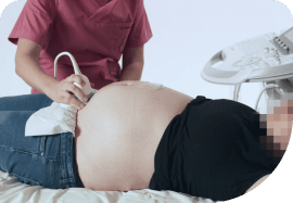
OB/GYN Ultrasound BachelorClass
7 CME credits
Basics of gynecology & obstetrics
instead of 840 €
only 588 €
Ultrasound Guided Regional Anaesthesia
Expertise in regional anaesthesia techniques
instead of 700 €
only 490 €
Pediatric Ultrasound BachelorClass
4.75 CME credits
Diagnose pediatric patients through ultrasound
instead of 770 €
only 539 €
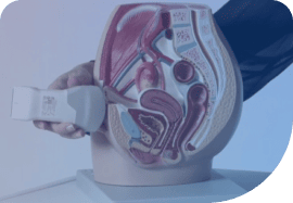
OB/GYN POCUS FocusClass
2 CME credits
Knowledge and skills on how to do an ultrasound examination in OB/GYN
instead of 420 €
only 294 €
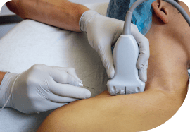
MSK ultrasound courses
Musculoskeletal Ultrasound BachelorClass
19.75 CME credits
Foundations of MSK ultrasound
instead of 990 €
only 693 €
MSK Guided Injections US MasterClass
4.75 CME credits
Advanced MSK ultrasound
instead of 770 €
only 539 €
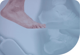
MSK Foot & Ankle Ultrasound BachelorClass
11.5 CME credits
Basics of MSK US of the foot and ankle joint
instead of 700 €
only 490 €
Bone Fracture FocusClass
2.5 CME credits
Ultrasound knowledge and skills specific to fracture sonography
instead of 300 €
only 210 €
Neuromuscular US Essentials
8.5 CME credits
Diagnose conditions and injuries not visible through other imaging methods
instead of 630 €
only 441 €
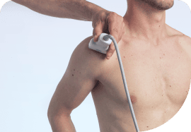
MSK Ultrasound of the Shoulder Joint
20 European CME credits
Ultrasound knowledge and skills specific to shoulder joint sonography
instead of 820 €
only 574 €

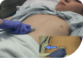
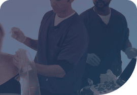
Emergency and Point-of-Care Ultrasound courses
Be prepared for emergencies at all times by untangling your ultrasound knowledge! Click on the arrows or swipe to see more courses!
Point-of-Care US FocusClass
6 CME credits
Primary care ultrasound
instead of 830 €
only 581 €
Emergency & Critical Care US Essentials
Essentials of emergency ultrasound
instead of 790 €
only 553 €
Prehospital Point-of-Care Ultrasound
10 CME credits
POCUS in prehospital scenarios
instead of 1,030 €
only 721 €
Emergency US MasterClass
5.25 CME credits
Advanced emergency ultrasound
instead of 770 €
only 539 €

Emergency US BachelorClass
Basic emergency ultrasound
instead of 700 €
only 490 €
Why our users love us

Ahmed Benzoghli
Wasseem Rock

Kati Tonninger, Austria
"I was recommended 123sonography through a friend and became a fan. The quality of the images and films are amazing."

Steven Feldstein, USA
"The e-learning platform is quite simple to navigate through, and the quality of the videos and images is quite good."

Satish Govind, India
"The communication style is simple, uncomplicated, but extremely effective in the way various topics are covered and discussed."